$1,167.00
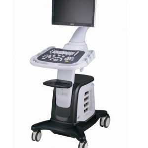
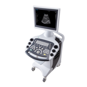
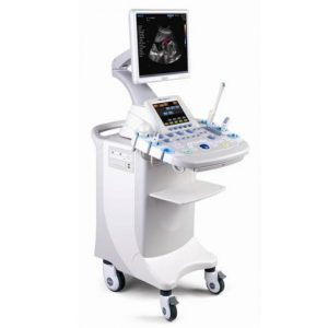
WED-180 is fully digital ultrasound system that is able to process real time image displays. It offers more than 40 body marks. Many probes are optional for clinic diagnosis demands. Offers high resolution.
Description
Ultrasound Scanner WED-180
Features
WED-180 adopts multiple technologies such as micro-computer control and digital scan converter (DSC), digital beam-forming (DBF), real time dynamic aperture (RDA), real time dynamic receiving apodization(DRA), real time Dynamic
receiving focusing (DRF), Digital frequency Scan (DFS) which can display a high quality, stable and high resolution image.
The external Video printer and external monitor display can be realized with the PAL-D Video output port. All the ultrasound
images can download to PC with high speed USB 2.0 port. Various image processing software packages are available.
This fully digital ultrasound system is used for abdominal, obstetric, cardiac and small body parts with ultrasound and sonography applications. Due to its compact in size and weight, it is very user-friendly.


|
Main Unit Parameters |
|
|
Scanning Model |
Electronic Linear Array, Electronic Convex Array |
|
Probe Connector |
2 |
|
Image Storage |
≥64 frames; external U Disk |
|
Display Depth |
250mm (maximum) |
|
Image Adjustment |
Black/White, Left/Right, Up/Down, Brightness, Scanning Line Density, Dynamic
Range, Focus Number, Focus Distance, Focus Position, Frame Correlation, M Speed and Sound Power |
|
Image Processing |
Image Smoothing/Sharpening, tissue harmonic, Gamma Correction, Histogram and Pseudo-color |
|
Measurement |
Distance, Perimeter, Area, Volume, Heart, GA, EDD, FW |
|
Report |
Automatically generate abdomen, urology, obstetrics and cardiac reports. |
Portable digital ultrasound system WED-180 / No Battery Included
CLINICAL APPLICATIONS:
- Abdominal.
- OB/GYN.
- Urology.
- Cardiology.
- Small parts.
FEATURES:
- Dynamic aperture technology to ensure image definition, from nearest to furthest fields.
- RDA: Real-time Dynamic Aperture.
- DFS: Dynamic Frecuency Scan.
- DRA: Dynamic Receiving Apodization.
- DRF: Dynamic Receiving Focusing.
- Creates records automatically (Normal/OB).
- Automatic probe identification (2 sockets).
- Backlit keypad.
- Digital controls.
- 8 segments TGC
TECHNICAL DATA:
- Scanning methods: Convex / Linear / Micro-Convex.
- Display modes: B, B+B, B+M, M, 4B, B+2B, 6B, 12B.
- Grayscale: 256.
- Scan depth: 40mm – 240 mm.
- CINE: >500 frames.
- Permanent image saving: 64 frames.
- Image flip: Up/down, left/right, black/ white
- Pseudo-Colour: 3.
- Zoom: 2 steps. Both in real time and frozen image.
- Measurements: Distance, Circunference, Area, Volume, Heart Rate.
- OB measurements: EDD, GA, FW (BPD, GS, CRL, FL, HC, AC).
- Ports: PAL-D, USB 2.0, RS-232.
- Display: LCD display 12.1″ (adjustable brightness/contrast).
- Body marks: 40.
- Comments: Date/Time, Name, Age, Gender, Doctor, Hospital, Notes.
- Power supply: AC 100V-240V, 50/60Hz.
STANDARD CONFIGURATION:
- Main unit.
- 2 probe sockets.
- Convex Probe
OPTIONAL PROBES:
- Ref. 369-PROBE-R20: Micro-Convex probe 5.0 MHz.
- Ref. 369-PROBE-EL: Linear Endorectal probe 7.5 MHz.
- Ref. 369-PROBE-HL: Linear probe 7.5 MHz.
- Ref. 369-PROBE-R13: Convex Transvaginal probe 6.5 MHz.
- Ref. 369-PROBE-R60: Convex probe 3.5 MHz.